photo sharing and upload picture albums photo forums search pictures popular photos photography help login
Douglas Stucky | profile | all galleries >> Galleries >> Brian tree view | thumbnails | slideshow

Brian

Click on the thumbnails below to see all the images in each gallery.

GDD. Gnathodiaphyseal dysplasia. We call it Brian's disorder.

February, 1996. Brian's sinus cavities beneath his eyes began to swell from what we thought was a sinus infection. His pediatrician prescribed antibiotics but the swelling didn't go down. After trying two more antibiotics that had no effect, he finally ordered a catscan.

The scan revealed that tumors were growing and a biopsy was ordered. After waiting a couple of long, long weeks for a diagnosis it was determined that Brian had a very rare bone disorder named polyostotic fibrous dysplasia.

A nurse from Children's Hospital asked us if we were familiar with The Magic Foundation, a group formed by other parents with children diagnosed with rare disorders. She gave us their contact information and we learned of a medical convention they were holding in Chicago. We drove to Chicago and learned a lot about fibrous dysplasia (FD) and met many families in the same situation as us...trying to learn more about what was going on in our childrens bodies.

At the convention, we met Dr. Collins from the National Institute of Health in Bethesda, MD. Dr. Collins was conducting a 3 year study of FD and after inquiring about Brian's case he said Brian would be an ideal candidate for the study. After much discussion, Carol and I decided to enroll Brian in the study with the hope of doing our small part to help find a cure for future generations.

In early December of 1999, Mr. Brian went to Washington. After a week of tests in which they weighed, measured, x-rayed, catscanned, photographed, took numerous blood and urine samples, tested for everything from scurvy to you name it, we were sent home to once again await the results. Before we left, Dr. Collins did remark that he felt that there was about a 70% chance that he had FD. What else could he have? At that point he didn't have an answer.

I think it was in February of 2000 that Dr. Collins called and said they had ruled out FD. He still didn't know what was causing the problems but said many team members were working on a diagnosis. Tissue samples were sent all over the world to pathologists trying to find a match to Brian's.
But nothing matched.

The exact date escapes me, but Dr. Collins called again to say they have finally given Brian's disorder a name. A detailed report was written to be published in The Journal of Bone and Mineral research where they proposed the name of this previously unknown disorder.

The following is from the report.

"Abstract

We report an unusual generalized skeletal syndrome characterized by fibro-osseous lesions of the jawbones with a prominent psammomatoid body component, bone fragility, and bowing/sclerosis of tubular bones. The case fits with the emerging profile of a distinct syndrome with similarities to previously reported cases, some with an autosomal dominant inheritance and others sporadic. We suggest that the syndrome be named gnathodiaphyseal dysplasia. The patient had been diagnosed previously with polyostotic fibrous dysplasia (PFD) elsewhere, but further clinical evaluation, histopathological study, and mutation analysis excluded this diagnosis. In addition to providing a novel observation of an as yet poorly characterized syndrome, the case illustrates the need for stringent diagnostic criteria for FD. The jaw lesions showed fibro-osseous features with the histopathological characteristics of cemento-ossifying fibroma, psammomatoid variant. This case emphasizes that the boundaries between genuine GNAS1 mutation-positive FD and other fibro-osseous lesions occurring in the jawbones should be kept sharply defined, contrary to a prevailing tendency in the literature. A detailed pathological study revealed previously unreported features of cemento-ossifying fibroma, including the participation of myofibroblasts and the occurrence of psammomatoid bodies and aberrant mineralization, within the walls of blood vessels. Transplantation of stromal cells grown from the lesion into immunocompromised mice resulted in a close mimicry of the native lesion, including the sporadic formation of psammomatoid bodies, suggesting an intrinsic abnormality of bone-forming cells.

Case History

The patient, a 5-year-old white boy, was enrolled in an ongoing study on the natural history of PFD conducted at the National Institutes of Health (NIH protocol 98-D-0145) after a diagnosis of PFD was made elsewhere. He was the child of nonconsanguineous parents with no family history of genetic or skeletal diseases or neurofibromatosis.

He presented at the age of 13 months with bilateral, relatively symmetric, expansile lesions in the maxillary bones associated with a sinus infection. Radiographic examinations at various ages and of various anatomical sites are shown (Fig. 1). In addition to their remarkable symmetry and expansile nature, the maxillary lesions exhibited a degree of lucency with occasional mineralized areas. A complete radiographic survey showed the additional findings of lytic lesions to the right and left of the midline in the mandible. Bowing and cortical thickening of the diaphyseal region of the tibias and fibulae were present. Similar changes also were observed in the right ulna and radius. The spine, femora, and growth plates all appeared normal. A bone scan revealed increased uptake in the maxilla and mandible, normal tracer distribution at the growth plates, and no uptake in the skull base or the tibias.

Incisional biopsy specimens of the right and left maxilla and mandible were initially read as consistent with FD. Subsequent review of the histological material at two major referral centers before enrolling in the NIH study concurred with the original reading of FD. At the age of 14 months, surgical reduction of maxillary lesions was performed, with removal of abnormal tissue and grafting of autologous iliac bone. A similar procedure was subsequently performed for treatment of the mandibular lesions, followed by three further surgeries on the maxillary lesions over the ensuing 3 years.

Over the same time interval, the patient sustained five fractures, all occurring as a result of minimal trauma. Sites of fracture included the left tibia and fibula, both clavicles (three fractures), and the left wrist.

Physical examination at the NIH revealed the symmetrical prominence of the cheeks bilaterally, and bowing of the distal lower extremities, left greater than right. The height and weight were normal for age. He was Tanner stage I and the rest of the physical examination was normal. Specifically, there was no evidence of café-au-lait skin pigmentation, neurofibromas, or Lisch nodules. Sclerae were white and erupted teeth were normal. There was no evidence of scoliosis, hypermobility of the joints, or arched palate. Neither the facial bone lesions nor the lower extremity lesions were painful to palpation.

Journal of Bone and Mineral Research, Journal of Bone and Mineral Research September 2001:16:1710-1718 (doi: 10.1359/jbmr.2001.16.9.1710)"

Here is the link to the article:

http://csdb.nidcr.nih.gov/papers/riminucci2001.pdf

And now, Brian is creating the history and prognosis of this disorder.

Brian's disorder. Brian's disease.

Brian's Battle.

Thanks for your prayers,

Doug and Carol


A CarePage has been set-up for Brian: http://www.carepages.com/
First time visitors must register before visiting Brian's CarePage. After you sign up, type in the name, briansbattle, where indicated.
IMG_1002crop.editir.jpg
Brian: The Early Years
gallery: Brian: The Early Years
Brian 2005
gallery: Brian 2005
Brian 2006
gallery: Brian 2006
Brian 2007
gallery: Brian 2007
Brian's Deck....A gift from The Powell Church...
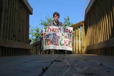
gallery: Brian's Deck....A gift from The Powell Church...
Brian 2008
gallery: Brian 2008
Brian 2009
gallery: Brian 2009
Brian 2010
gallery: Brian 2010
Brian 2011
gallery: Brian 2011
Brian 2012
gallery: Brian 2012
Brian...in Memorium
gallery: Brian...in Memorium